

How Have Animals Contributed To Help In The T-celll Therape
Introduction
Great advances in immunology are unremarkably supported by experiments carried out in animal models and by far, inbred lines of mice and their respective knock-out or knock-in derivatives, are the virtually commonly used fauna systems in immunological studies. Though with much credit to their usefulness, laboratory mice will never provide all the answers to fully understand immunological processes. Also, some answers provided in mouse models are non applicable to other species of animals or humans. Large animal models offer unique biological and experimental advantages that have been and continue to be of great value to the understanding of biological and immunological processes.
The humble cow, the underestimated pig and the unassuming chicken have greatly influenced our current understanding of human immunology. For about immunologists dedicated to fundamental and applied research, it is piece of cake to forget that B cells were offset identified in chickens and vaccination first occurred because of a cow. Although there are far too many important events to discuss in this paper, we have chosen to highlight a few of the most important contributions of farm animals to the current understanding of immunology (Table 1).
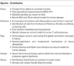
Tabular array 1. Selected major contributions of subcontract animals to immunology.
History of Vaccination
Edward Jenner published in 1798 a booklet entitled "An Inquiry into the Causes and Effects of the Variolae Vaccinae, a disease discovered in some of the western counties of England, particularly Gloucestershire and known by the name of Cow Pox" (1, 2) and although strictly speaking Jenner did non discover vaccination, he was the first person to utilise scientific rigor to evidence protection from disease through targeted intervention. The English dairy farmer Benjamin Jesty (1737–1816) was the offset person known to vaccinate confronting smallpox (iii) protecting his family against the virus fifty-fifty after numerous exposures (3).
However, the idea and indeed the term vaccination, only came into the light spot 100 years later thanks to Louis Pasteur. This fourth dimension the chicken takes a privileged position and the story was beautifully explained by Pasteur himself (four, 5) and has been romanticized in Paul De Kruif's volume "Microbe Hunters" (6). In 1878 Pasteur inoculated chickens with "stale" cultures of Pasteurella multocida. The chickens became ill but recovered then he decided to re-inoculate them with a fresh civilization. The chickens that had received the "stale" culture recovered whereas chickens that had not been pre-exposed to the stale cultures died. Pasteur recognized the similarities betwixt his studies in chickens and what Jenner had published with smallpox. He coined the term "vaccine" (four, 5, seven) in honor of Jenner.
By the early 1880s, William Smith Greenfield in the UK (eight, 9) and Pasteur working with Henri Thullier, Charles Chamberland and Pierre Paul Émile Roux in France (10, 11) had begun developing and testing vaccines confronting anthrax in sheep and cattle. A decade subsequently, the German scientists Friedrich Loffler and Paul Frosch identified the first ever filterable infectious agent in mammals: foot and rima oris disease virus (FMDV) and developed a fully protective heat-inactivated vaccine against it (12, 13); however an effective long-lasting and broadly protective vaccine against FMDV remains elusive.
Pigs also played an important role in early on vaccinology studies. By the late 1800s swine plague or hog cholera (later discovered to be caused by a virus now called classical swine fever virus, CSFV (xiv) was killing hundreds of thousands of pigs across the give-and-take and was particularly of business organisation to the Usa pig producing manufacture, causing an impressive US$15 million a year in losses in 1875 (fifteen) and United states of america$xx million past 1878one. In one case again, Pasteur and Thullier developed a vaccine to what is at present thought to be the start ever vaccine against a viral infectious disease (sixteen) and the starting time mass-vaccination campaign in history. In improver, information technology is rarely recognized that CSFV was the start animal virus ever to be cultured in vitro (17) and the techniques developed past Carl Tenbroek keep to be used today.
Horses accept besides contributed to the agreement of fundamental immunological mechanisms. In a serial of experiments, Emile Roux working with Alexandre Yersin and followed past Emil von Behring and Shibasaburo Kitasato immunized horses to produce an "antidote" or allowed sera against the diphtheria toxin that was somewhen used to care for humans, an important step in understanding antibodies and humoral immunity (eighteen). Behring won the Nobel Prize for Medicine in 1901 for this work.
Another milestone in vaccine development was the generation in the 1970's of vaccines to control Marek'south disease (Dr.), a naturally occurring neoplastic disease in chickens acquired past an oncogenic herpesvirus (xix). Md vaccines are the start examples of the use of vaccination to protect confronting cancer (xx, 21).
With the discovery of molecular biology techniques in the 1960'south and 70'south, the race was on to develop recombinant vaccines against numerous infectious diseases. The first report of a biosynthesized polypeptide vaccine was published in 1981 (22). The structural protein VP3 of FMDV was cloned and expressed in E. coli and the purified protein used to vaccinate six cattle and two swine, which developed neutralizing antibodies and were protected against claiming with FMDV (22). And new technologies take only helped to highlight the importance of farm animals in vaccine development: using a computational arroyo to assess protein-poly peptide stability, Kotecha and colleagues (23) used molecular dynamic ranking to predict FMDV capsid stabilities and produced stabilized FMDV capsids based on these predictions, assessed their stability using X-ray crystallography and demonstrated their improved immunogenicity in vivo by vaccinating cattle. This demonstrates the potential value of structure-based blueprint of vaccines to develop stabilized vaccine antigens for animals and humans alike.
Innate Immunity
Although the innate immune organization of animals is largely conserved, there are significant variations in the Blueprint-Recognition-Receptor (PRR) structures of various species (24). It has been suggested that laboratory mice have not been subjected to the selective pressures that other animals have and and so innate immune studies carried out in laboratory animals do not accurately inform human being biology (25). It has been demonstrated that homo and subcontract fauna PRR responses to their ligands (24, 26) are more similar to each other than homo-mouse PRR responses (26–28). Considering PRR recognition is associated with adaptive amnesty, a better understanding of these molecules in farm animals is likely to better inform on their effect in these animals as well as humans.
A major contribution of the chicken to fundamental innate immunity was the description in 1957 of the offset interferon. Chicken embryos were exposed to influenza virus by Alick Issacs and Jean Lindenmann (29) and they identified an allowed soluble element responsible for regulating virus infection. This discovery was certainly i of the scientific landmarks in cell biology in the Twentieth century and one which opened the doors of what we now know as innate immunity.
B Cells
Perhaps the most recognizable contribution of the chicken to science, and immunology in particular, was in the definition of the 2 elements of adaptive immunity: the B-dependent and the T-dependent immunity.
The avian bursa of Fabricius, named afterward Hieronymus Fabricius of Aquapendente (30) is a sac-similar construction located in the cloacal passage of the bird and its function remained elusive until well into the Twentieth century. Bruce Glick and Timothy Chang working at the Poultry Science Section at Ohio State University (30, 31) described how post-obit the surgical removal of the bursa, chickens injected with Salmonella typhimurium "O" antigen failed to develop bacteria-specific antibodies. Glick and Chang wrote a newspaper entitled: "The role of the bursa of Fabricius in antibody production" and was rejected past leading scientific journals (xxx) and eventually published in Poultry Science (32). Several years later, the bone marrow in mammals was shown to be the equivalent of the bursa in birds (33), and then the term "B-lymphocyte" originated from "bursa-derived lymphocyte". Several years later, Cooper et al. published a central paper on the demarcation of the thymic and bursal and systems in birds and proposed the beingness of equivalent systems in mammals (34).
The cannulation of lymphatic vessels was adult in the early Twentieth century in rats to study the lymphatic system but due to the complication of the surgical process, sheep and and so cattle were used extensively in the 1960s and 70s in lymphatic cannulation studies (35). In a series of adoptive transfers of lymph-migrating cells and fluid, Hall and colleagues offset identified in sheep that antibody-secreting cells (ACS) run across antigens and are activated in the lymph nodes (36), then drift via the efferent lymphatics to the circulatory system, and that the immune response depends on an intact lymphatic system.
Cattle have also contributed to fundamental B cell immunology and the generation of a highly diverse antibody repertoire. Most vertebrates encode a large number of variable (V), diversity (D), and joining (J) gene segments and antibody variety is achieved by recombination of these 3 segments. In contrast, cattle only express a limited number of V genes and so it is thought that antibody diverseness is achieved recombination events and endogenous mutation mechanisms in the CDR3 region (37). Another unusual feature of bovine antibodies is their exceptionally long CDR3 regions (38). These long CDR3 and unusual mutation mechanisms effect in "microfolds" within the CDR3 region that allow bovine antibodies to demark antigens that would normally be inaccessible (38).
A recent report demonstrated that cows can be immunized with a single HIV Env trimer and this results in strong HIV-specific nAbs which are dependent on the length of the CDR3 loops of bovine Ig (39). It has been suggested that this could be an efficient style of producing super-antibodies against other human being pathogens. Transchromosomal cows have been engineered to produce large amounts of full human IgG molecules with pathogen specificity: MERS-CoV (40), Hanta virus (41), VEEV (42), and Ebola virus (43). This technology has the potential to generate prophylactic antibodies against emerging viral diseases.
On the other hand, chickens have serum IgM and IgA both of which are homologs of their mammalian counterparts; in improver, they limited IgY, not found in mammals but idea to be an evolutionary ancestor for mammalian IgG and IgE. Chickens however do non have either IgE nor IgD but instead apply a distinct process for generating antibody diversity that is distinctly different to mammals (44).
Engineers frequently wait to nature for inspiration. Antibody engineers are no exception, modeling new therapeutics on molecules found in animals such every bit camels and cows. Indeed, ten% of bovine antibodies have unusually long heavy-chain CDR3s as part of their antigen-recognition sites. Stanfield et al. (45) take solved crystal structures of three new bovine Fab fragments and analyzed the five known structures to testify that their ultra-long CDR H3s all prefer like architectures composed of a knob domain containing a small conserved β-sheet connected past diverse disulfide-bonded loops that is separated from the antibody surface past a long conserved stalk. They propose that varying the length of the stalk and the positions and number of disulphide links in the knob may help drive antibody variety. These structural insights could be leveraged to tailor antibiotic-based therapeutics.
In contrast to all other mammals, camelids (dromedaries, camels, llamas, etc) also take an unique antibody type similar IgG but with identical heavy chains lacking the CH1 domain and which do non pair with their corresponding lite chains. These "heavy-chain antibodies" (HCAbs) display antigen-specific variable domains or "VHH" which are structurally and functionally like to an IgG Fv but have simply three CDR loops defining the antigen biding sites. Camel VHH domains, besides called "nanobodies," have been of swell interest because of their stability and pocket-size size and stiff affinity to their corresponding antigens. In fact, several camel VHH domain antibodies are in early preclinical development in oncology, infectious, inflammatory, and neurodegenerative diseases (44), the most contempo instance being the generation of broadly neutralizing antibodies to influenza in llamas (46).
T Cells
Cytotoxic and helper T cells are generally considered to be phenotypically different due to the mutually exclusive expression of the co-receptors CD8αβ and CD4 and differences in MHC-restriction (class I vs. class II). Withal, between 3 (in normal individuals) and lx% (in sure pathologies) of human peripheral blood lymphocytes take been shown to be CD4/CD8 double positive (DP) T cells (47). Thymic and extra-thymic development of T cells has been studied mainly in mice and considering the expression of CD8 and CD4 in mouse T cells for the well-nigh part mutually exclusive, CD4/CD8 DP lymphocytes accept generally been ignored. Nevertheless, the presence of CD4/CD8 DP T cells in many animals makes it impossible to ignore these cells. Studies in pigs have shown that CD4/CD8 DP are a singled-out subset of activated and/or memory T helper cells (48) and in humans the increment in circulating CD4/CD8 DP T cell frequency has been identified in autoimmune and chronic inflammatory diseases (49–53) suggesting the importance of this particular T prison cell population in human health.
Most circulating T cells in humans and mice are conventional T cells expressing the αβ T jail cell receptor (TCR) and either CD4 or CD8. Dissimilar mice, other species like cattle, pigs and chickens possess a substantial proportion of T cells expressing the γδ TCR cells in the circulation suggesting that circulating γδ TCR T cells have a more than of import role immunity than previously thought (54).
The fact that the phenotype and frequency of circulating and tissue resident T cells is then vastly inconsistent in different species suggests that immune responses to (vaccine) antigens are also distinct. Information technology is assumed that that all animal species have a similar immune response to a particular antigen, but this is a statement to exist reviewed in light of each host particularities. In addition, the TH1/TH2 polarization of T cells observed in response to particular antigens is a phenomenon of certain strains of laboratory mice and not of outbred mammals including subcontract animals and humans (55, 56). In fact, it has been shown that cytokine profiles defining TH1/TH2 responses to antigens in cattle are more than like to human responses than those observed in mice (57).
Dendritic Cells
Dendritic cells (DC) equally such, and their role in amnesty were beginning described in the 1970s and in 1995 Ralph Steinman published a series of papers describing that a cellular receptor called "December-205" (now CD205) was expressed on mouse DC, was involved in antigen processing (58, 59) and was detected by the monoclonal antibody NLDC-145. In fact, it was 2 years earlier in 1993, that Chris Howard, a bovine immunologist working at the then called "Institute for Animal Heath" in the United kingdom published a series of papers identifying an important and until then uncharacterized marker expressed on pseudo-afferent lymph veiled cells (too chosen ALDC) detected past the monoclonal antibody WC9 (now CC98) (60–63). Although Steinman'southward identification of mouse CD205 helped characterize the binding of CC98 to bovine CD205 (64), the importance of CD205 in identifying DC was beginning axiomatic in cattle.
As mentioned above, Steinman's seminal work in characterizing DC using the mouse organisation has been one of the most important developments in cellular immunology of the Twentieth century, and ane which lead to his Nobel laureate. Withal, the idea that a component of the immune organization was involved in antigen processing and presentation had been proposed many times before. Every bit mentioned to a higher place, cannulation of the lymphatic vessels is more than applied in large than small animals, and this technique has been used to investigate DC biology. Afferent or peripheral lymph DC were first described in sheep in 1972 (65) equally "very phagocytic dendritic macrophages that are involved in long term immunological reactions" that are very strong antigen presenting non-lymphoid cells (66) and that their phagocytic and antigen presentation capacities differed from "classical" peritoneal macrophages (67), therefore indicating that DC and macrophages were dissimilar cell types (67) several years earlier Steinman'due south observations (68). In improver, lymphatic cannulation of sheep has revealed important ontologic, phenotypic and functional characteristics of DC subsets that are relevant in other mammals, particularly humans (69, seventy).
Similitudes and differences betwixt swine and human DC/macrophage populations have recently been described (71). In 1 striking instance and in contrast to studies performed in mice, swine and human cDC2, which are associated with Th2 responses, both limited FcεRIα and are localized in or next to the tracheal and bronchial epithelia. These observations have been proposed to imply that swine and humans accept similar allergen responses as opposed to mice. This theory is supported by the fact that localization of cDC2 helps them access antigens such every bit airborne allergens, and FcεRIα expression on these cells might help proliferation observed in allergic responses.
γδ T Cells
As mentioned before and dissimilar mice, horses and humans, near other animals have a large γδ T cell compartment. For example, up to 70% of all blood lymphocytes in young calves are T cell expressing the γδ T jail cell receptor (TCR). Although the reasons for the enlarged T prison cell compartment in cattle, pigs and chickens is all the same unknown, their large numbers and ease of collection has resulted in bang-up advances in γδ T cell biology knowledge not just for farm animals, just also for humans. For instance, APCs were shown to influence γδ T cell proliferation (72, 73). Cynthia Baldwin's lab has divers antigen-specific bovine γδ T cell responses in various systems (74–76) and Adrian Smith's lab has washed similar observations in chickens (77, 78). Information technology has too been shown that bovine γδ T are potent regulatory T cells (79), an ascertainment that is also true for a subset of human γδ T cells (80, 81). These results in farm animals have and continue to enhance our understanding of homo γδ T biological science (82). Perhaps the most important one was the realization that a subset of bovine γδ T cells expressed MHC class II and co-stimulatory molecules on their surface, a characteristic normally attributed to macrophages, B cells and DC just not T cells (83). Bovine γδ T cells were also shown to phagocyte antigens and of MHC 2-restricted presentation to CD4+ T cells (83). This function of bovine γδ T cells was later on reported in pigs (84) and much subsequently in mouse (85) and human (86–88) γδ T cells.
Somatic Cell Nuclear Transfer
Perhaps the best known contribution of whatever farm brute to scientific progress was the somatic cell nuclear transfer that gave origin to Dolly, the sheep (89). Although nuclear transfer itself is not a direct contribution to immunology, nuclear transfer technology has directly influenced many immunological concepts underpinned past technologies such as induced pluripotent stem (iPS) cells and CRISPR-Cas systems.
Clustered regularly interspaced short palindromic repeats (CRISPR) is a RNA-guided endonuclease used both in vivo and in vitro. Genetically modified animals becoming more common and their availability can be exploited in many applications such every bit comparative immunology, physiology and disease, to generate in vivo bioreactors to produce complex proteins, or to produce genetically modified organs for transplantation in humans (90).
Fauna Models of Infection and Affliction
Although the majority of pharmaceutical enquiry is performed in laboratory mice models, it is clear that humans are not "large mice." By a big extent, studies in laboratory mice take been the victim of over interpretation; for example, by extrapolating successful pre-treatment in mice to therapeutic handling in men. The weakness of the mouse model in pharmaceutical research was recently highlighted in a study showing that inflammatory responses in mouse models do non correlate with homo inflammatory disease (91). An boosted study showed a close similarity in expression profiles of immune-related genes between humans and pigs (92).
Cattle, pigs, and chickens, are useful, valid, and valuable models to study homo infectious diseases and of import clinical targets in their own right. Both humans and cattle are the natural hosts for tuberculosis (being infected with the genetically-related Mycobacterium bovis and Mycobacterium tuberculosis, respectively) and the bovine and human diseases share many similarities in terms of immunity and pathology (93), whereas the mouse model of tuberculosis does not provide a true-blue representation of the disease in humans (94). Similarly, bovine respiratory syncytial virus (bRSV) is closely related to human (h) RSV and the pulmonary pathology, immune responses and epidemiology seen in immature calves and children are very similar (95). Swine have been shown to be a more faithful model for human influenza infection and amnesty studies and the aforementioned strains of influenza infect both humans and pigs considering the distribution of flu virus receptors and physiopathology are similar in both species. The transfer of maternal-derived antibodies (MDA) to new born pigs enables fancy vaccine written report design to elucidate the role of MDA in immunity (96, 97), vaccine efficacy and in enhancement of respiratory disease (98).
Gnotobiotic piglets have been used to study diverse human gastrointestinal pathogens. For instance, human noroviruses are antigenically and genetically related to swine noroviruses and unlike mice, humans and pigs testify genetic susceptibilities to noroviruses depending on their histoblood group antigen phenotypes and the virus strain. Similarly, gnotobiotic pigs have been used in rotavirus research to study disease pathogenesis and place virus-specific IgA and ASC as correlates for protection and vaccine efficacy in children (99). Pigs accept also been proposed to be better models than mice for many other infectious diseases including female genital infection with Chlamydia trachomatis, Helycobacter pylori, Neisseria meningitides, and Nipah virus amid others considering of the natural susceptibility of pigs to these pathogens (100).
Endogenous retroviruses were starting time discovered in pig kidney prison cell lines in 1971 (101) and are now known to be present in nearly, if non all, mammalians. The presence and potential reactivation of endogenous retroviruses has very important consequences in both allo- and xeno-transplantation.
Immunotherapy is becoming more than pop in clinical trials and vaccine efficacy studies. The success of allowed cell therapies partially depends on the constructive delivery of cells to target organs, a process that invariably involves the lymphatic system. DC migration in mice has non proven to exist very informative, however, DC migration in pigs may be able to answer several question on DC migration that cannot be addressed otherwise. These studies demonstrate that using big animals to investigate immune cell trafficking volition aid improve immunotherapies in humans (102).
Xenotransplantation
In 1906 the French surgeon Mathieu Jaboulay (1860–1913) implanted a pig'south kidney into one woman and a goat's liver into some other thus starting the idea of xenotransplantation; unsurprisingly, both women died (103). The acceptance or rejection of a donor'southward organ or cells is fundamentally an immunological event. Cellular rejection involves NK and T cells that recognize foreign antigens on the grafted tissue. Using xenotransplantation models (pig-to-rat, pig-to-primate, and pig-to-homo), the main mechanism for organ and tissue rejection has been proposed to involve arteriosclerosis, or thickening of the arterial walls. This procedure if thought to be caused past activated and allo-reactive lymphocytes that migrate over time to the transplanted organ (104). Arteriosclerosis is a major cause of chronic organ rejection (103).
Developmental Immunology
Studies in laboratory mice have underpinned many concepts of allowed tolerance and the generation of immune responses in the neonate. Yet, the peripheral immune arrangement in mice remains unpopulated during pregnancy and information technology is only after nascence that B and T cells brainstorm to emmigrate to the periphery. In dissimilarity, lymphoid cells circulate through the fetus in humans and large animals well-before nascence and specialized lymphoid tissues are besides well-developed and populated past the fourth dimension of birth and are able to answer to a number of antigens (105, 106). Certainly the allowed system in neonate humans and large animals is not matured, but calves, lambs and piglets can be more than useful than mice in understanding immune responses during pregnancy and in new borns and these studies can exist used to better inform man developmental immunology. This advantage over mice has recently been used to develop extracorporeal support technologies using neonatal lambs with the ultimate objective to utilise these technologies in premature children (107).
Large Animal Models in Vaccinology
Perhaps one of the most common uses of large animal models is in the development of vaccines with several advantages over mice. The serial drove of peripheral blood from animals such as pigs, cattle, chickens, and horses allows for immunokinetic studies to exist possible in response to vaccination or infection at the level of the individual. These immunokinetic studies can be used to correlate immune responses generated with protection after challenge with the relevant pathogen. In vaccinology studies using mice, the typical approach would be to sacrifice groups of mice sequentially and harvest spleen and claret, then the immune response to vaccination at the private level is not unremarkably achieved.
Large animals also provide several advantages over mice when investigating mucosal amnesty. When mice are vaccinated or inoculated intranasally, it is common for the inoculum to exist digested because anesthetized mice can both eat and inhale the fabric placed on their olfactory organ. In addition, the structure of the mucosal associated lymphoid tissue (MALT) differs significantly in mice from that of large animals and humans; for instance mice practise not accept tonsils but instead have undefined networks of MALT, whereas cattle, pigs and sheep have well-defined tonsils (108–110). In the case of vaccine delivery through the skin, cattle, and pigs appear to be improve suited than mice for these studies. Skin thickness, structure of the epidermis and the presence and distribution of Langerhan'south cells are amid many characteristics that humans and cattle and pigs accept in mutual and which are practically relevant in transcutaneous immunizations (111).
Limitations of Animal Models
The process of selecting a relevant and appropriate creature model is a counterbalanced and complicated exercise due to the diversity in vertebrate physiology, adaptive and innate immunity. Studies in mice, for instance, accept shown the efficacy of vaccines against FMDV, even so these efficacy studies have failed to be translated to the target species (cattle and pigs), presumably due to fundamental differences in the immune systems of model organisms and target species and the ability of the virus to mutate in these animals (112). Information technology has recently been shown that because immunoglobulin subclass diversification occurred after speciation (113, 114) a particular immunoglobulin subclass in one species bears no functional homology to ane of the same name of another species (115). Thus, our knowledge of the functions of IgG1 in mice cannot be extrapolated to other mammals. Characterizing generating reagents for each animal model hinders the evolution and usefulness of whatever of these models and therefore limiting the usefulness of cows, cattle or chickens as models for human immunology.
Mice and rats are and will probably go on to be the chosen model organisms over subcontract animals. Mice tin be readily mutated (knock in or knock out) to study immunological pathways; so far this has been proven to be very difficult—and expensive—in large animals. As mentioned above, the availability of reagents to study immune cells and processes in mice far out competes the availability of these reagents for large animals. Pharmacokinetic and toxicology studies would be prohibitively expensive in pigs, horses or cattle, so small-scale rodents and rabbits are the best organisms to use in these studies. In addition, studies in mice have been primal in the discovery of antibiotics, chemotherapy agents and more recently Auto-T jail cell therapies that tin be directly applied to humans. Genetic homogeneity, low cost, the availability of biologically-relevant mutants and reagents make the mouse the optimal brute model for many academic and industrial researchers.
Conclusions
Subcontract animals have historically contributed and continue to contribute to fundamental and applied immunology. The use of these animals in research is not difficult every bit long every bit the appropriate facilities and reagents are bachelor. Dedicated housing, cost, biosecurity, and genetic variability are some of the many disadvantages confronted when using farm animals in enquiry. However, selecting an appropriate creature model should be more than just a matter of accessibility and mutual practice (116) but should be based on the scientific question to be addressed and its relevance.
Author Contributions
All authors listed have made a substantial, straight and intellectual contribution to the piece of work, and approved information technology for publication.
Funding
The authors were funded by the UK's BBSRC grants Bulletin board system/E/I/00002067 and BBS/Eastward/I/00002014.
Conflict of Interest Statement
The authors declare that the research was conducted in the absence of any commercial or fiscal relationships that could exist construed as a potential conflict of interest.
Footnotes
one. ^(1881). Swine Plague. Scientific discipline 2, 121
References
four. Pasteur. On the germ theory. Science (1881) two:420–2. doi: x.1126/science.os-2.63.420
CrossRef Full Text
v. Pasteur ML. On craven cholera: written report of the conditions of not-recidivation and of some other characteristics of this disease. Science (1881) ii:126–7. doi: 10.1126/scientific discipline.os-two.39.126
CrossRef Full Text | Google Scholar
8. Greenfield WS. Lectures on some recent investigations into the pathology of infective and contagious diseases. Lancet (1880) 115:865–vii. doi: ten.1016/S0140-6736(02)42734-1
CrossRef Full Text | Google Scholar
9. Tigertt WD. Anthrax. William Smith Greenfield, M.D., F.R.C.P., Professor Superintendent, the Brown Animal Sanatory Establishment (1878-81). Apropos the priority due to him for the production of the first vaccine against anthrax. J Hyg. (1980) 85:415–twenty. doi: 10.1017/S0022172400063488
PubMed Abstruse | CrossRef Full Text | Google Scholar
11. Pasteur L, Chamberland Roux. Summary report of the experiments conducted at Pouilly-le-Fort, most Melun, on the anthrax vaccination, 1881. Yale J Biol Med. (2002) 75:59–62.
PubMed Abstract | Google Scholar
12. Loeffler FAF, Paul. Summarischer Bericht uber dice Ergebnisse der Untersuchungen der Kommission zur Erforschung der Maul- und Klauenseuche bei dem Institut fur Infektionskrankheiten in Berlin. Centralblatt fur Bakteriologie, Parasitenkunde und Infektionskrankheiten, Abt I (1897) 22:257–9.
13. Loeffler FAF, Paul. Berichte der Kommission zur Erforschung der Maul- und Klauenseuche bei dem Institut fur Infektionskrankheiten in Berlin. Centralblatt fuXr Bakteriologie, Parasitenkunde und Infektionskrankheiten, Abt I (1898) 23:371–91.
xv. Partridge JM. Swine plague, or pig cholera. Public Health Pap Rep. (1883) 9:347–51.
Google Scholar
16. Pasteur L, Thuillier L, Frank RM, Wrotnowska D. Correspondence of Pasteur and Thuillier Concerning Anthrax and Swine Fever Vaccinations. Tuscaloosa, AL: Universit Ala, Academy of Alabama Printing (1969).
19. Witter RL, Moulthrop JI, Burgoyne GH, Connell HC. Studies on the epidemiology of Marek's illness herpesvirus in broiler flocks. Avian Dis. (1970) 14:255–67. doi: 10.2307/1588470
PubMed Abstract | CrossRef Full Text | Google Scholar
xx. Purchase HG, Okazaki Due west. Consequence of vaccination with herpesvirus of turkeys (HVT) on horizontal spread of Marek'southward affliction herpesvirus. Avian Dis. (1971) fifteen:391–7. doi: 10.2307/1588710
PubMed Abstract | CrossRef Full Text | Google Scholar
22. Kleid DG, Yansura D, Small B, Dowbenko D, Moore DM, Grubman MJ, et al. Cloned viral poly peptide vaccine for foot-and-oral cavity affliction: responses in cattle and swine. Scientific discipline (1981) 214:1125–ix. doi: x.1126/scientific discipline.6272395
PubMed Abstract | CrossRef Full Text | Google Scholar
23. Kotecha A, Seago J, Scott K, Burman A, Loureiro S, Ren J, et al. Structure-based energetics of poly peptide interfaces guides human foot-and-mouth disease virus vaccine design. Nat Struct Mol Biol. (2015) 22:788–94. doi: 10.1038/nsmb.3096
PubMed Abstract | CrossRef Full Text | Google Scholar
25. Werling D, Jann OC, Offord V, Drinking glass EJ, Coffey TJ. Variation matters: TLR construction and species-specific pathogen recognition. Trends Immunol. (2009) 30:124–thirty. doi: x.1016/j.it.2008.12.001
PubMed Abstract | CrossRef Full Text | Google Scholar
26. Grabiec A, Meng G, Fichte Southward, Bessler W, Wagner H, Kirschning CJ. Human simply not murine toll-like receptor 2 discriminates betwixt tri-palmitoylated and tri-lauroylated peptides. J Biol Chem. (2004) 279:48004–12. doi: x.1074/jbc.M405311200
CrossRef Total Text | Google Scholar
28. Farhat Thou, Riekenberg S, Jung Thousand, Wiesmuller KH, Jungi TW, Ulmer AJ. Identification of full length bovine TLR1 and functional characterization of lipopeptide recognition past bovine TLR2/1 heterodimer. Vet Res. (2010) 41:34. doi: 10.1051/vetres/2010006
PubMed Abstract | CrossRef Full Text | Google Scholar
29. Isaacs A, Lindenmann J. Virus interference. I The interferon. Proc R Soc Lond B Biol Sci. (1957) 147:258–67. doi: 10.1098/rspb.1957.0048
CrossRef Total Text | Google Scholar
30. Taylor RL Jr, McCorkle FM Jr. A landmark contribution to poultry scientific discipline–Immunological office of the bursa of Fabricius. Poult Sci. (2009) 88:816–23. doi: 10.3382/ps.2008-00528
PubMed Abstract | CrossRef Full Text | Google Scholar
31. Glick B. Growth of the bursa of fabricius and its relationship to the adrenal gland in the white pekin duck, white leghorn, outbred new-hampshire, and inbred new-hampshire. Poultry Sci. (1959) 38:1208–1208.
32. Glick B, Chang TS, Jaap RG. The bursa of fabricius and antibody product. Poultry Sci. (1956) 35:224–5. doi: 10.3382/ps.0350224
CrossRef Full Text | Google Scholar
33. Becker AJ, McCulloch EA, Till JE. Cytological demonstration of the clonal nature of spleen colonies derived from transplanted mouse marrow cells. Nature (1963) 197:452–4. doi: 10.1038/197452a0
PubMed Abstruse | CrossRef Full Text | Google Scholar
35. Lascelles AK, Morris B. Surgical techniques for the collection of lymph from unanaesthetized sheep. Q J Exp Physiol Cogn Med Sci. (1961) 46:199–205. doi: 10.1113/expphysiol.1961.sp001536
PubMed Abstract | CrossRef Total Text | Google Scholar
36. Hall JG, Morris B, Moreno GD, Bessis MC. The ultrastructure and function of the cells in lymph post-obit antigenic stimulation. J Exp Med. (1967) 125:91–110. doi: 10.1084/jem.125.ane.91
PubMed Abstract | CrossRef Full Text | Google Scholar
37. Zhuang Y, Futse JE, Brown WC, Brayton KA, Palmer GH. Maintenance of antibody to pathogen epitopes generated by segmental gene conversion is highly dynamic during long-term persistent infection. Infect Immun. (2007) 75:5185–90. doi: 10.1128/IAI.00913-07
PubMed Abstract | CrossRef Total Text | Google Scholar
39. Sok D, Le KM, Vadnais K, Saye-Francisco KL, Jardine JG, Torres ZL, et al. Rapid elicitation of broadly neutralizing antibodies to HIV by immunization in cows. Nature (2017) 548:108–11. doi: 10.1038/nature23301
PubMed Abstract | CrossRef Full Text | Google Scholar
40. Luke T, Wu H, Zhao J, Channappanavar R, Coleman CM, Jiao JA, et al. Human polyclonal immunoglobulin Grand from transchromosomic bovines inhibits MERS-CoV in vivo. Sci Transl Med. (2016) eight:326ra321. doi: 10.1126/scitranslmed.aaf1061
PubMed Abstract | CrossRef Full Text | Google Scholar
41. Hooper JW, Brocato RL, Kwilas SA, Hammerbeck CD, Josleyn MD, Royals M, et al. DNA vaccine-derived human IgG produced in transchromosomal bovines protect in lethal models of hantavirus pulmonary syndrome. Sci Transl Med. (2014) 6:264ra162. doi: x.1126/scitranslmed.3010082
PubMed Abstract | CrossRef Full Text | Google Scholar
42. Gardner CL, Sun C, Luke T, Raviprakash G, Wu H, Jiao JA, et al. Antibody preparations from human transchromosomic cows exhibit safety and therapeutic efficacy against venezuelan equine encephalitis virus. J Virol. (2017) 91:e00226–17. doi: 10.1128/JVI.00226-17
PubMed Abstract | CrossRef Full Text | Google Scholar
43. Dye JM, Wu H, Hooper JW, Khurana S, Kuehne AI, Coyle EM, et al. Product of potent fully human polyclonal antibodies against ebola zaire virus in transchromosomal cattle. Sci Rep. (2016) 6: 24897. doi: 10.1038/srep24897
PubMed Abstruse | CrossRef Full Text | Google Scholar
44. de los Rios M, Criscitiello MF, Smider VV. Structural and genetic diversity in antibody repertoires from diverse species. Curr Opin Struct Biol. (2015) 33:27–41. doi: 10.1016/j.sbi.2015.06.002
CrossRef Full Text | Google Scholar
45. Stanfield RL, Wilson IA, Smider VV. Conservation and diversity in the ultralong third heavy-chain complementarity-determining region of bovine antibodies. Sci Immunol. (2016) ane:aaf7962. doi: 10.1126/sciimmunol.aaf7962
CrossRef Total Text | Google Scholar
46. Laursen NS, Friesen RHE, Zhu X, Jongeneelen Yard, Blokland Southward, Vermond J, et al. Universal protection against influenza infection by a multidomain antibody to influenza hemagglutinin. Scientific discipline (2018) 362:598–602. doi: 10.1126/science.aaq0620
PubMed Abstract | CrossRef Full Text | Google Scholar
47. Blue ML, Daley JF, Levine H, Schlossman SF. Coexpression of T4 and T8 on peripheral claret T cells demonstrated by two-color fluorescence catamenia cytometry. J Immunol. (1985) 134:2281–6.
PubMed Abstract | Google Scholar
48. Talker SC, Kaser T, Reutner Chiliad, Sedlak C, Mair KH, Koinig H, et al. Phenotypic maturation of porcine NK- and T-prison cell subsets. Dev Comp Immunol. (2013) 40:51–68. doi: 10.1016/j.dci.2013.01.003
PubMed Abstract | CrossRef Full Text | Google Scholar
49. Berrih Due south, Gaud C, Bach MA, Le Brigand H, Binet JP, Bach JF. Evaluation of T cell subsets in myasthenia gravis using anti-T cell monoclonal antibodies. Clin Exp Immunol. (1981) 45:1–viii.
PubMed Abstract | Google Scholar
50. Munschauer FE, Stewart C, Jacobs 50, Kaba South, Ghorishi Z, Greenberg S, et al. Circulating CD3+ CD4+ CD8+ T lymphocytes in multiple sclerosis. J Clin Immunol. (1993) xiii:113–8. doi: 10.1007/BF00919267
PubMed Abstract | CrossRef Full Text | Google Scholar
53. Quandt D, Rothe Yard, Scholz R, Baerwald CW, Wagner U. Peripheral CD4CD8 double positive T cells with a distinct helper cytokine profile are increased in rheumatoid arthritis. PLoS ONE (2014) 9:e93293. doi: 10.1371/periodical.pone.0093293
PubMed Abstract | CrossRef Full Text | Google Scholar
54. Takamatsu HH, Denyer MS, Stirling C, Cox S, Aggarwal Northward, Dash P, et al. Porcine gammadelta T cells: possible roles on the innate and adaptive immune responses following virus infection. Vet Immunol Immunopathol. (2006). 112:49–61. doi: 10.1016/j.vetimm.2006.03.011
PubMed Abstruse | CrossRef Full Text | Google Scholar
55. Dicks MD, Guzman East, Spencer AJ, Gilbert SC, Charleston B, Hill AV, et al. The relative magnitude of transgene-specific adaptive immune responses induced by human and chimpanzee adenovirus vectors differs between laboratory animals and a target species. Vaccine (2015) 33:1121–8. doi: 10.1016/j.vaccine.2015.01.042
PubMed Abstract | CrossRef Full Text | Google Scholar
56. Mendonca SC. Differences in immune responses confronting Leishmania induced by infection and past immunization with killed parasite antigen: implications for vaccine discovery. Parasit Vectors (2016) nine:492. doi: ten.1186/s13071-016-1777-10
PubMed Abstract | CrossRef Full Text | Google Scholar
58. Jiang W, Swiggard WJ, Heufler C, Peng M, Mirza A, Steinman RM, et al. The receptor DEC-205 expressed by dendritic cells and thymic epithelial cells is involved in antigen processing. Nature (1995) 375:151–5. doi: 10.1038/375151a0
PubMed Abstract | CrossRef Full Text | Google Scholar
59. Swiggard WJ, Mirza A, Nussenzweig MC, Steinman RM. Dec-205, a 205-kDa protein abundant on mouse dendritic cells and thymic epithelium that is detected by the monoclonal antibiotic NLDC-145: purification, label, and N-concluding amino acid sequence. Cell Immunol. (1995) 165:302–xi. doi: 10.1006/cimm.1995.1218
PubMed Abstruse | CrossRef Full Text | Google Scholar
61. Naessens J, Howard CJ. Leukocyte antigens of cattle and sheep. Monoclonal antibodies submitted to the 2d Workshop. Vet Immunol Immunopathol. (1993) 39:5–10. doi: 10.1016/0165-2427(93)90157-Y
CrossRef Full Text | Google Scholar
62. Parsons KR, Bembridge G, Sopp P, Howard CJ. Studies of monoclonal antibodies identifying two novel bovine lymphocyte antigen differentiation clusters: workshop clusters (WC) 6 and seven. Vet Immunol Immunopathol. (1993) 39:187–92. doi: 10.1016/0165-2427(93)90180-C
PubMed Abstract | CrossRef Full Text | Google Scholar
63. Howard CJ, Sopp P, Brownlie J, Parsons KR, Lee LS. Phenotypic variation and functional differences within dendritic cells isolated from afferent lymph. Adv Exp Med Biol. (1995) 378:105–7. doi: 10.1007/978-i-4615-1971-3_22
PubMed Abstruse | CrossRef Full Text | Google Scholar
64. Gliddon DR, Promise JC, Brooke GP, Howard CJ. DEC-205 expression on migrating dendritic cells in afferent lymph. Immunology (2004) 111:262–72. doi: 10.1111/j.0019-2805.2004.01820.x
PubMed Abstract | CrossRef Full Text | Google Scholar
65. Morris B. The Cells of Lymph and Their Office in Immunological Reactions. Handbuch der Allgemeinen Pathologie. Berlin: Springer Verlag. (1972) 3:405. doi: x.1007/978-3-642-51133-2_10
CrossRef Full Text
66. Emery DL, MacHugh ND, Ellis JA. The properties and functional activity of non-lymphoid cells from bovine afferent (peripheral) lymph. Immunology (1987) 62:177–83.
PubMed Abstract | Google Scholar
67. Barfoot R, Denham South, Gyure LA, Hall JG, Hobbs SM, Jackson LE, et al. Some backdrop of dendritic macrophages from peripheral lymph. Immunology (1989) 68:233–9.
PubMed Abstract | Google Scholar
68. Steinman RM, Cohn ZA. Identification of a novel cell blazon in peripheral lymphoid organs of mice. Morphology, I., quantitation, tissue distribution. J Exp Med. (1973) 137:1142–62. doi: 10.1084/jem.137.5.1142
PubMed Abstract | CrossRef Full Text | Google Scholar
69. Pascale F, Contreras V, Bonneau Yard, Courbet A, Chilmonczyk South, Bevilacqua C, et al. Plasmacytoid dendritic cells drift in afferent skin lymph. J Immunol. (2008) 180:5963–72. doi: ten.4049/jimmunol.180.nine.5963
PubMed Abstract | CrossRef Full Text | Google Scholar
seventy. Contreras V, Urien C, Guiton R, Alexandre Y, Vu Manh TP, Andrieu T, et al. Existence of CD8alpha-like dendritic cells with a conserved functional specialization and a mutual molecular signature in distant mammalian species. J Immunol. (2010) 185:3313–25. doi: x.4049/jimmunol.1000824
PubMed Abstract | CrossRef Full Text | Google Scholar
71. Maisonnasse P, Bouguyon E, Piton G, Ezquerra A, Urien C, Deloizy C, et al. The respiratory DC/macrophage network at steady-state and upon influenza infection in the swine biomedical model. Mucosal Immunol. (2016) 9:835–49. doi: 10.1038/mi.2015.105
PubMed Abstruse | CrossRef Total Text | Google Scholar
72. Okragly AJ, Hanby-Flarida M, Baldwin CL. Monocytes control gamma/delta T-cell responses by a secreted product. Immunology (1995) 86:599–605.
PubMed Abstract | Google Scholar
73. Okragly AJ, Hanby-Flarida Thousand, Mann D, Baldwin CL. Bovine gamma/delta T-jail cell proliferation is associated with self-derived molecules constitutively expressed in vivo on mononuclear phagocytes. Immunology (1996) 87:71–ix.
PubMed Abstract | Google Scholar
74. Naiman BM, Alt D, Bolin CA, Zuerner R, Baldwin CL. Protective killed Leptospira borgpetersenii vaccine induces potent Th1 immunity comprising responses past CD4 and gammadelta T lymphocytes. Infect Immun. (2001) 69:7550–viii. doi: x.1128/IAI.69.12.7550-7558.2001
PubMed Abstract | CrossRef Total Text | Google Scholar
75. Baldwin C, Hsu H, Chen C, Palmer M, McGill J, Waters WR, et al. The role of bovine γδ T cells and their WC1 co-receptor in response to bacterial pathogens and promoting vaccine efficacy: a model for cattle and humans. Vet Immunol Immunopathol. (2014) 159:144–55. doi: 10.1016/j.vetimm.2014.02.011
PubMed Abstract | CrossRef Full Text | Google Scholar
76. Baldwin CL, Telfer JC. The bovine model for elucidating the role of γδ T cells in decision-making infectious diseases of importance to cattle and humans. Mol Immunol. (2015) 66:35–47. doi: x.1016/j.molimm.2014.10.024
PubMed Abstract | CrossRef Full Text | Google Scholar
77. Smith AL, Hayday AC. An alphabeta T-prison cell-contained immunoprotective response towards gut coccidia is supported by gammadelta cells. Immunology (2000) 101:325–32. doi: 10.1046/j.1365-2567.2000.00122.x
PubMed Abstract | CrossRef Full Text | Google Scholar
78. Inagaki-Ohara G, Sakamoto Y, Dohi T, Smith AL. γδ T cells play a protective part during infection with Nippostrongylus brasiliensis by promoting goblet prison cell function in the small intestine. Immunology (2011) 134:448–58. doi: 10.1111/j.1365-2567.2011.03503.x
PubMed Abstract | CrossRef Total Text | Google Scholar
79. Guzman E, Hope J, Taylor G, Smith AL, Cubillos-Zapata C, Charleston B. Bovine γδ T cells are a major regulatory T cell subset. J Immunol. (2014) 193:208–22. doi: ten.4049/jimmunol.1303398
PubMed Abstract | CrossRef Total Text | Google Scholar
80. Huang Y, Aydintug MK, Loomis J, Macleod MK, McKee AS, Kirchenbaum 1000, et al. Antigen-specific regulation of IgE antibodies past not-antigen-specific γδ T cells. J Immunol. (2013) 190:913–21. doi: ten.4049/jimmunol.1202230
PubMed Abstract | CrossRef Full Text | Google Scholar
81. Mao Y, Yin S, Zhang J, Hu Y, Huang B, Cui L, et al. A new event of IL-4 on human γδ T cells: promoting regulatory Vδ1 T cells via IL-x production and inhibiting office of Vδ2 T cells. Prison cell Mol Immunol. (2016) 13:217–28. doi: x.1038/cmi.2015.07
PubMed Abstract | CrossRef Full Text | Google Scholar
82. Guzman East, Price S, Poulsom H, Promise J. Bovine γδ T cells: cells with multiple functions and important roles in immunity. Vet Immunol Immunopathol. (2012) 148:161–67. doi: x.1016/j.vetimm.2011.03.013
PubMed Abstruse | CrossRef Total Text | Google Scholar
83. Collins RA, Werling D, Duggan SE, Banal AP, Parsons KR, Howard CJ. Gammadelta T cells present antigen to CD4+ alphabeta T cells. J Leukoc Biol. (1998) 63:707–fourteen. doi: 10.1002/jlb.63.6.707
PubMed Abstract | CrossRef Full Text
84. Takamatsu HH, Denyer MS, Wileman TE. A sub-population of circulating porcine gammadelta T cells tin human activity as professional person antigen presenting cells. Vet Immunol Immunopathol. (2002) 87:223–4. doi: x.1016/S0165-2427(02)00083-i
PubMed Abstruse | CrossRef Full Text | Google Scholar
85. Cheng L, Cui Y, Shao H, Han Thousand, Zhu L, Huang Y, et al. Mouse gammadelta T cells are capable of expressing MHC form 2 molecules, and of functioning equally antigen-presenting cells. J Neuroimmunol. (2008) 203:three–11. doi: 10.1016/j.jneuroim.2008.06.007
PubMed Abstract | CrossRef Full Text | Google Scholar
87. Brandes M, Willimann K, Bioley One thousand, Levy Northward, Eberl M, Luo One thousand, et al. Cross-presenting human gammadelta T cells induce robust CD8+ alphabeta T cell responses. Proc Natl Acad Sci United states of america. (2009) 106:2307–12. doi: 10.1073/pnas.0810059106
PubMed Abstract | CrossRef Total Text
88. Meuter S, Eberl Thousand, Moser B. Prolonged antigen survival and cytosolic consign in cantankerous-presenting human gammadelta T cells. Proc Natl Acad Sci USA. (2010) 107:8730–five. doi: ten.1073/pnas.1002769107
PubMed Abstract | CrossRef Total Text | Google Scholar
90. Lamas-Toranzo I, Guerrero-Sanchez J, Miralles-Bover H, Alegre-Cid G, Pericuesta E, Bermejo-Alvarez P. CRISPR is knocking on befouled door. Reprod Domest Anim. (2017) 52 (Suppl. 4):39–47. doi: 10.1111/rda.13047
PubMed Abstract | CrossRef Total Text | Google Scholar
91. Seok J, Warren HS, Cuenca AG, Mindrinos MN, Baker HV, Xu W, et al. Genomic responses in mouse models poorly mimic human inflammatory diseases. Proc Natl Acad Sci The states. (2013) 110:3507–12. doi: 10.1073/pnas.1222878110
PubMed Abstract | CrossRef Full Text | Google Scholar
92. Dawson HD, Loveland JE, Pascal G, Gilbert JG, Uenishi H, Isle of man KM, et al. Structural and functional annotation of the porcine immunome. BMC Genomics (2013) xiv:332. doi: ten.1186/1471-2164-xiv-332
PubMed Abstract | CrossRef Full Text | Google Scholar
93. Waters WR, Palmer MV, Thacker TC, Davis WC, Sreevatsan South, Coussens P, et al. Tuberculosis immunity: opportunities from studies with cattle. Clin Dev Immunol. (2011). 2011:768542. doi: x.1155/2011/768542
PubMed Abstract | CrossRef Full Text | Google Scholar
94. Gupta UD, Katoch VM. Beast models of tuberculosis for vaccine development. Indian J Med Res. (2009) 129:11–8. doi: 10.4103/ijmr.IJMR_554_18
PubMed Abstract | CrossRef Full Text
96. Renshaw HW. Influence of antibody-mediated allowed suppression on clinical, viral, and immune responses to swine influenza infection. Am J Vet Res. (1975) 36:v–xiii.
PubMed Abstract | Google Scholar
97. Genzow M, Goodell C, Kaiser TJ, Johnson Due west, Eichmeyer Thousand. Live attenuated influenza virus vaccine reduces virus shedding of newborn piglets in the presence of maternal antibody. Influenza Other Respir Viruses (2017) 12:353–9. doi: 10.1111/irv.12531
PubMed Abstract | CrossRef Full Text | Google Scholar
98. Rajao DS, Perez DR. Universal vaccines and vaccine platforms to protect against influenza viruses in humans and agriculture. Forepart Microbiol. (2018) 9:123. doi: x.3389/fmicb.2018.00123
PubMed Abstract | CrossRef Total Text | Google Scholar
99. Saif LJ, Ward LA, Yuan Fifty, Rosen BI, To TL. The gnotobiotic piglet equally a model for studies of affliction pathogenesis and immunity to human rotaviruses. Arch Virol Suppl. (1996) 12:153–61. doi: 10.1007/978-three-7091-6553-9_17
PubMed Abstract | CrossRef Full Text | Google Scholar
102. Crisci E, Fraile 50, Novellas R, Espada Y, Cabezon R, Martinez J, et al. In vivo tracking and immunological backdrop of pulsed porcine monocyte-derived dendritic cells. Mol Immunol. (2015) 63:343–54. doi: x.1016/j.molimm.2014.08.006
PubMed Abstract | CrossRef Full Text | Google Scholar
104. Groth CG, Korsgren O, Wennberg Fifty, Tibell A, Zhu Due south, Sundberg B, et al. Xenoislet rejection following pig-to-rat, pig-to-primate, and pig-to-man transplantation. Transplant Proc. (1996) 28:538–9.
PubMed Abstruse | Google Scholar
106. Mutwiri G, Bateman C, Baca-Estrada ME, Snider Yard, Griebel P. Induction of immune responses in newborn lambs post-obit enteric immunization with a human adenovirus vaccine vector. Vaccine (2000) 19:1284–93. doi: ten.1016/S0264-410X(00)00230-9
PubMed Abstruse | CrossRef Full Text | Google Scholar
107. Partridge EA, Davey MG, Hornick MA, McGovern PE, Mejaddam AY, Vrecenak JD, et al. An extra-uterine system to physiologically support the extreme premature lamb. Nat Commun. (2017) 8:15112. doi: 10.1038/ncomms15112
CrossRef Full Text | Google Scholar
108. Mestas J, Hughes CC. Of mice and not men: differences between mouse and human immunology. J Immunol. (2004) 172:2731–8. doi: 10.4049/jimmunol.172.5.2731
CrossRef Full Text | Google Scholar
109. Garraud O, Borhis G, Badr G, Degrelle S, Pozzetto B, Cognasse F, et al. Revisiting the B-cell compartment in mouse and humans: more one B-cell subset exists in the marginal zone and beyond. BMC Immunol. (2012) 13:63. doi: x.1186/1471-2172-13-63
PubMed Abstract | CrossRef Total Text | Google Scholar
111. Hammond SA, Tsonis C, Sellins K, Rushlow K, Scharton-Kersten T, Colditz I, et al. Transcutaneous immunization of domestic animals: opportunities and challenges. Adv Drug Deliv Rev. (2000) 43:45–55. doi: 10.1016/S0169-409X(00)00076-4
PubMed Abstract | CrossRef Total Text | Google Scholar
112. Habiela M, Seago J, Perez-Martin Eastward, Waters R, Windsor M, Salguero FJ, et al. Laboratory animal models to study human foot-and-mouth disease: a review with emphasis on natural and vaccine-induced amnesty. J Gen Virol. (2014) 95(Pt eleven):2329–45. doi: ten.1099/vir.0.068270-0
PubMed Abstract | CrossRef Full Text | Google Scholar
113. Butler JE, Weber P, Wertz N. Antibiotic repertoire evolution in fetal and neonatal piglets. XIII Hybrid VH genes and the preimmune repertoire revisited. J Immunol. (2006) 177:5459–70. doi: x.4049/jimmunol.177.8.5459
CrossRef Full Text | Google Scholar
114. Qin T, Ren Fifty, Hu X, Guo Y, Fei J, Zhu Q, et al. Genomic organization of the immunoglobulin calorie-free chain gene loci in Xenopus tropicalis: evolutionary implications. Dev Comp Immunol. (2008) 32:156–65. doi: 10.1016/j.dci.2007.05.007
PubMed Abstract | CrossRef Total Text | Google Scholar
115. Morgan SB, Holzer B, Hemmink JD, Salguero FJ, Schwartz JC, Agatic One thousand, et al. Therapeutic administration of broadly neutralizing FI6 antibiotic reveals lack of interaction between human IgG1 and Pig Fc receptors. Forepart Immunol. (2018) 9:865. doi: 10.3389/fimmu.2018.00865
PubMed Abstruse | CrossRef Full Text | Google Scholar
Source: https://www.frontiersin.org/articles/10.3389/fvets.2018.00307/full
Posted by: baileycoluch.blogspot.com
0 Response to "How Have Animals Contributed To Help In The T-celll Therape"
Post a Comment